A 52-year-old male alcoholic patient with hypertension and a prior history of brain ischemia was admitted to the neurosurgery department with signs and symptoms of ischemic stroke. A transesophageal echocardiogram and computed tomography suggested a possible pedunculated thrombus at the transition of the ascending aorta to the aortic arch. The therapeutic option was surgical treatment using cardiopulmonary bypass, deep hyperthermia and total cardiac arrest. The procedure involved resection of the aortic wall together with the thrombus to avoid recurrence. There were no adverse consequences and the patient was released from hospital with the use of oral anticoagulant drugs.
Homem, 52 anos de idade, alcoólatra, hipertenso, com história prévia de isquemia cerebral. Admitido no Departamento de Neurocirurgia com sinais e sintomas de acidente vascular cerebral isquêmico. A ecocardiografia transesofágica e tomografia computadorizada evidenciaram imagem sugestiva de trombo pedunculado ao nível da transição entre a aorta ascendente e o arco aórtico. O tratamento de escolha foi procedimento cirúrgico com emprego da circulação extracorpórea associada a hipotermia profunda e parada circulatória total. Foi realizada ressecção do trombo e de parte parede da aorta para evitar reincidência. O paciente não apresentou intercorrências e teve alta hospitalar com uso de anticoagulante oral.
INTRODUCTION
The presence of an atheroma plaque existent in the intimal layer of the aorta gives an unknown potential for the formation of non-obstructive pedunculated thrombi, which is a rare alteration as the layers of the aorta can be normal.
Thromboembolic phenomena in this situation are frequent complications. BARBON GARCIA et al. reported occlusion of the retinal artery arising from a pedunculated thrombus located in the aortic arch. [1] Involvement of arteries of lower limbs and coronary arteries can expose the patient to catastrophic consequences of peripheral arterial insufficiency and acute myocardial infarction, respectively. [2,3]
Recently, RIBEIRO et al. published a report of the surgical treatment of a non-occlusive pedunculated thrombus of the ascending aorta in a patient with prior clinical history of strokes. The objective of this case report is to show the risks of extracardiac thromboembolic events.
CASE REPORT
A 52-year-old male patient with history of alcoholism, hypertension and three episodes of strokes in a period of four years is reported. The physical examination demonstrated the presence of hemiplegia on the right, a reduced pulse in the left femoral artery, dyslalia and bilateral loss of vision.
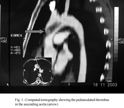
A transesophageal echocardiogram showed an image suggestive of a pedunculated thrombus at the junction of the ascending aorta and the aortic arch. Visualization was difficult due to the distance of the aorta from the esophagus. Computed tomography confirmed the diagnosis and made it possible to measure the size of the thrombotic mass (Fig. 1 - tomography).
For the operative procedure, cardiopulmonary bypass circulation was utilized via the femoral artery associated with deep hypothermia, total circulatory arrest and microcardioplegia with myocardial protection. The approach was through the anterior wall of ascendant aorta with the incision extending to the middle third of the aortic arch. The floating pedunculated thrombus that was adhered to the anterior wall of the aorta was identified and resected. A section of the aorta where the thrombus was adhered was incised and subsequently reconstruction of the aorta using bovine pericardium was performed (Figure 2).

The site of the adherence of the thrombus in the aorta was apparently, an atheroma plaque and the wall of the adjacent vessel had a normal appearance. A macroscopic histopathologic examination confirmed it to be a firm elastic, yellow membranous fragment measuring 2.8 x 2.7 cm and on one of its faces there was a friable, black, polypoid formation measuring 4.l x 2.4 x l.2 cm. By microscopy a fragment of elastic artery with accentuated alterations due to atherosclerosis on the tunica intima and tunica media was evidenced, with an area of erosion in continuity with a pedunculated formation constituted by alternating strips of erythrocytes and fibrin.
The postoperative evolved without adverse events and the patient was discharged from hospital on the seventh postoperative day using oral anticoagulants.
COMMENTS
The presence of pedunculated thrombi in arterial channels with high pressure and blood flow is rare however their complications are serious owing to the possibility of recurring thromboembolic events marked by extracardiac or cardiac sequels [2].
Transesophageal echocardiography is the best diagnostic method to identify a pedunculated thrombus in the thoracic aorta and to detect minimum intimal ulcerative injuries. During surgery echocardiography serves as a guide for the surgeon [5]. Some difficulties to clearly visualize the origin and extension of the thrombotic mass can occur. For this, magnetic resonance or computed tomography should be performed, mainly in cases in which the thrombus originates at the junction of the ascending aorta and the aorta arch.
Intravenous heparinization followed by oral anticoagulants is one clinical therapeutic alternative indicated for patients with a high risk to the operative procedure. Stollberger et al. in 2001, demonstrated absorption of the thrombus after 10 weeks and absence of recurrence over 30 months of following up [6].
Cardiopulmonary bypass associated with deep hyperthermia and total circulatory arrest was necessary for the approach and assessment of the aorta arch. It is fundamental that resection of the aorta wall adjacent to the thrombus is extensive to avoid recurrence. In spite of frequent and serious complications and that this seems to be a high-risk disease, the choice of surgery has proven to be effective and safe.
BIBLIOGRAPHIC REFERENCES
1. Barbon Garcia JJ, Rodriguez Blanco V, Alvarez Suarez ML, Fernandez Lombardia M, Diez-Lage Sanchez A. A retinal embolism subsequent to atheroma in aortic arch. Arch Soc Esp Oftalmol 2001; 76:735-8.
[ Medline ]
2. Lozano P, Gomez FT, Julia J, M-Rimbau E, Garcia F. Recurrent embolism caused by floating thrombus in the thoracic aorta. Ann Vasc Surg 1998; 12:609-11.
[ Medline ]
3. Bruno P, Massetti M, Babatasi G, Khayat A. Catastrophic consequences of a free floating thrombus in ascending aorta. Eur J Cardiothorac Surg 2001; 19:99-101.
[ Medline ]
4. Ribeiro PJF, Menardi AC, Vicente WVA, Évora PRB. Tratamento cirúrgico de um trombo pedunculado não oclusivo da aorta ascendente: apresentação de caso. Rev Bras Cir Cardiovasc 2003; 18:73-6.
[ SciELO ]
5. Gabelmann M, Geibel A, Redecker M, Fraedrich G, Just H. The aortic arch as source of thromboembolism events: significance of echocardiography diagnosis. Z Kardiol 1995; 84:729-32.
[ Medline ]
6. Stollberger C, Kopsa W, Finsterer J. Resolution of an aortic thrombus under anticoagulant therapy. Eur J Cardiothorac Surg 2001; 20:880-2.
[ Medline ]
 All scientific articles published at www.bjcvs.org are licensed under a Creative Commons license


 Read in Portuguese
 Portuguese PDF
 Print
Print
 Send this article by email
 How to cite this article
 Submit a comment
 Mendeley
 Pocket